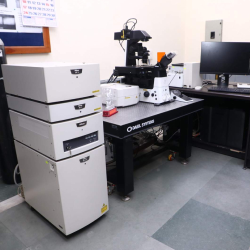
Confocal Microscope
Requisition forms
Please download the requisition form, fill it in, sign it, and upload the filled copy with your booking. This requisition form is provided by the USIC office for this instrument.
Process: submit booking → in-charge approval → pay on the DU fee portal → upload Order ID + receipt → measurement confirmed. See the payment flowchart and user guidelines.
About this instrument
Model- Eclipse Ti2 A1R
FEATURES
- Live-cell imaging
- High-speed time-lapse studies
- Super-resolution microscopy
- Quantitative fluorescence analysis
- 3D and 2D tracking system for particle tracking.
- Fluorescence Recovery after Photo bleaching (FRAP), Förster Resonance Energy
- Transfer (FRET), photo activation, and radiometric calcium imaging.
- DE convolution software for improved visualization
SPECIFICATION
Scanning Modes: Galvano scanner for standard imaging and resonant scanner for
high-speed imaging up to 230 frames per second (512 × 64 pixels).
Laser Lines: Equipped with multiline laser (405/488/561/647 nm).
Detectors: Standard fluorescence detector (400–750 nm) and Spectral detector (405–
650 nm).
Objectives: Supports high-resolution objectives, such as PLAN APOCHROMAT VC
20X N.A. 0.75 and PLAN APOCHROMAT VC 60X WI N.A. 1.2, achieving lateral
resolutions down to 0.18 µm.Software: Integrated with NIS-Elements C software for comprehensive image
acquisition.
SAMPLE- Slide Prepared